
Our investigation into iron metabolism and overload has revealed the critical function of the liver as the main storage site for iron. However, this raises an important question: How can we accurately assess the level of iron stored in the liver without relying on invasive techniques?
Assessing Liver Iron Levels
Historically, liver iron levels have been determined through liver biopsy, a method that involves taking a small sample of liver tissue for chemical examination. While this approach is indeed effective, it is invasive, uncomfortable, and not suited for repeated assessments. Additionally, the procedure comes with its own set of risks and drawbacks, making it far from ideal for routine clinical application.
Fortunately, advances in medical technology have introduced non-invasive alternatives that leverage the magnetic properties of iron to offer reliable evaluations of liver iron levels. Two prominent techniques making strides in this area are superconducting Quantum interference device-based magnetic susceptometry (SQUID) and magnetic resonance imaging (MRI).
SQUID
SQUID is a physics-based technique that utilizes the magnetic attributes of iron to measure liver iron concentration. This method involves applying a powerful magnetic field to the liver and measuring signal intensity variations as the organ shifts away from the detector. Although SQUID has proven effective in quantifying liver iron content, its broader use is limited by the availability and accessibility of SQUID machines.
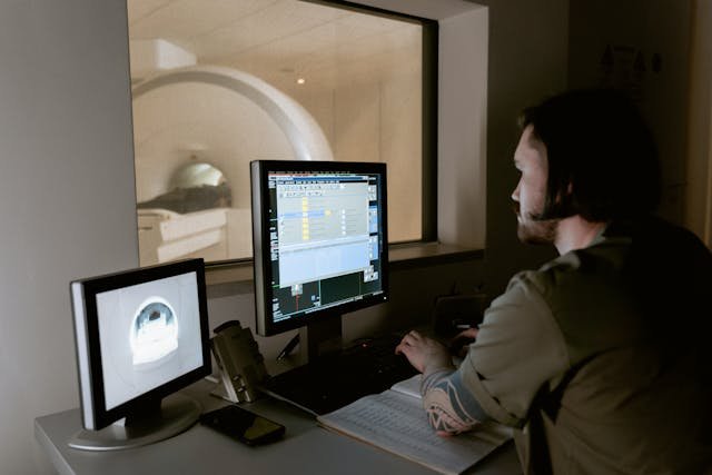
MRI
In contrast, MRI provides a more widely accessible and versatile approach to evaluating liver iron levels. MRI scanners are commonly found in hospitals and radiology centers, making them a practical option for non-invasive assessments. By utilizing the magnetic properties of iron particles within the liver, MRI can generate quantitative images that represent liver iron concentrations. This technique, known as FerriScan, uses water molecules in the tissue as conduits to detect the magnetic fields produced by iron particles. Employing advanced mathematical algorithms, FerriScan creates detailed maps of liver iron distribution, allowing clinicians to evaluate iron levels with greater accuracy than a biopsy.
The introduction of MRI technology has transformed the landscape of liver iron quantification, providing a safer, more comfortable, and more convenient alternative to traditional biopsy methods. With MRI, patients can undergo evaluations without the discomfort of invasive procedures or related recovery times, thereby enhancing their overall experience and adherence to diagnostic protocols.
In diagnosing hereditary hemochromatosis and other iron-related conditions, MRI plays a crucial role in identifying iron overload and guiding treatment options. When genetic tests fall short in confirming hereditary hemochromatosis, FerriScan offers a conclusive way to evaluate iron status, allowing healthcare providers to adopt tailored treatment plans that cater to individual patient requirements.
Additionally, FerriScan enables continuous monitoring of liver iron levels over time, offering important insights into treatment efficacy and disease progression. By tracking these iron levels longitudinally, healthcare professionals can refine patient care strategies, ensuring individuals with iron-related disorders receive timely interventions to minimize the complications associated with excess iron.
Conclusion
Innovative methods for assessing liver iron levels, including MRI-based techniques like FerriScan, signify a major advancement in the realm of iron metabolism and overload. By providing a non-invasive, precise, and easily accessible way to quantify liver iron concentration, FerriScan enhances our capacity to diagnose, monitor, and manage iron-related disorders effectively, ultimately fostering improved patient care and health outcomes.